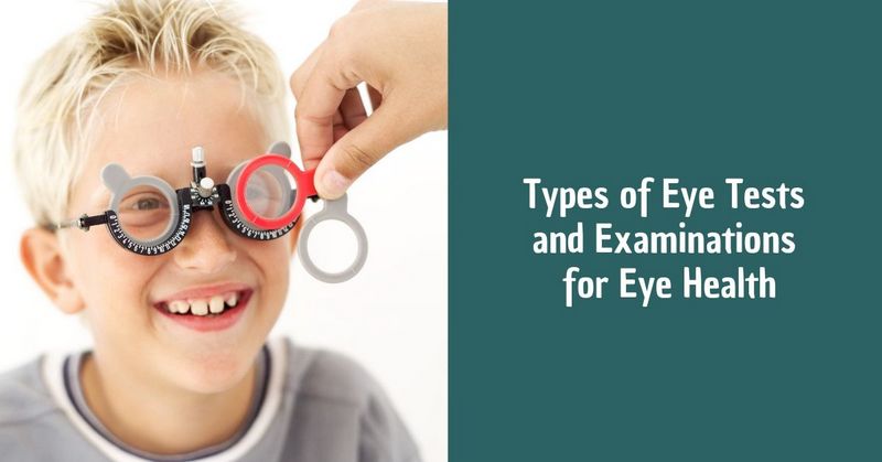
Types of Eye Tests and Examinations for Eye Health

Diagnosis of vision is a complex of accurate methods for detecting eye diseases. It helps to identify almost all pathologies, including the most minor deviations from the norm. Using computer diagnostics of vision, a specialist can make a complete and thorough examination of all structures of the eye and make an accurate diagnosis. Timely and correct diagnosis contributes to the choice of effective treatment. And this is VERY important because many eye diseases can be successfully cured or corrected if not delayed and done on the basis of an unmistakable diagnosis.
Tasks of eye exam diagnostics
Eye exams are performed to:
- assess the patient’s eye condition, identification diseases and pathologies at any stages;
- determine the best treatment program to prevent complications;
- establish the feasibility of surgical treatment;
- recognize an ailment before a person himself sees deterioration;
- identify signs of hidden diseases;
- diagnose possible serious illnesses;
- adjust age-related changes.
Types of an eye examination
The determination of visual fields is carried out using a contrasting study, which can help evaluate the approximate degree of loss of visual fields.
Evaluation of pupillary response to light (indirect and involuntary) allows an eye doctor to assess the condition of the optic tract. The absence of a direct light reflex is observed with unilateral damage to the optic nerve and occlusion of the central retinal artery.
Intraocular pressure is usually measured using a non-contact tonometer. If necessary, the measurement of intraocular pressure is carried out using a contact tonometer or a Goldman tonometer. To exclude glaucoma, it may be required to do computer perimetry, that is, the study of visual fields.
A refractive examination is performed before any surgical intervention. The exam includes determination of visual acuity with and without correction, biomicroscopy, ophthalmoscopy, tonometry, refractometry (using an auto refractometer), computer corneal topography on a computer topograph, ultrasound biometry, ultrasound pachymetry. The data obtained during diagnosis are used by the surgeon during excimer laser correction.
Before performing refractive surgery, patients undergo pachymetry with a corneal thickness measuring device, which allows calculating the maximum allowable depth of laser exposure, which in cases of a very high degree of myopia determines how much correction is possible.
Any microsurgical or laser interventions are preceded by a full comprehensive computer-aided diagnostic eye exams. The examination reveals the range of existing problems and determines the tactics of treatment.
Any eye disease requires a special approach in treatment, including at the stage of making a diagnosis. Many ophthalmic diseases have similar symptoms, so even experienced professionals cannot make a diagnosis and determine the degree of the disease without a thorough examination using specialized equipment. At an early stage, it is advisable to carry out diagnostics as planned. Below you can see a summary of the main methods for diagnosing eye diseases.
- A vision test through special tables and test lenses. Currently, their place is gradually occupied by more modern projectors and phoropters. This study is subjective since it largely depends on the state of the eye and the body at the time of the diagnosis. The procedure also involves the selection of spectacle lenses;
- Autorefractometry is a study of refraction and curvature of the eye cornea, performed using an automatic refracto keratometer. Using this device allows a doctor to fully automate the process. All that is required of the patient is to look at a special mark, thereby holding his or her head in a static position;
- Keratometry is the measurement of the corneal curvature. This study is used to calculate the optical power of an artificial lens and also allows detecting and measuring astigmatism. Keratometry is carried out using a special device, a keratometer;
- Tonometry is a measurement of intraocular pressure. It is used to diagnose glaucoma and disorders in the functioning of the optic nerve. The tonometry technique is simple: a specialist adds drops to the patient’s eye that reduces the sensitivity of the cornea and then applies an ophthalmic tonometer to its surface. The device exerts a slight pressure on the cornea, and the result of the study is its inverse resistance;
- Pachymetry is the process of measuring the thickness of the cornea. The study is carried out using ultrasound. It is used to diagnose corneal dystrophy. It is actively used before laser correction and to assess the condition of the eyes after corneal transplantation;
- Biomicroscopy is a visual examination of the cornea, lens, iris and other parts of the eye by creating a contrast between the illuminated and unlit areas. This allows an eye doctor to assess the condition of optical media, blood vessels and optic nerves;
- Ophthalmoscopy is the most common examination using a slit lamp, during which a doctor examines the eyeball and adnexa. It is carried out with the aim of diagnosing diseases of the cornea, iris, lens and vitreous body;
- Gonioscopy is an eye examination to look at the front part of your eye. It is performed using a slit lamp and gonioscope, a system of mirrors. It is carried out in order to detect pathologies of the anterior chamber angle (tumors, foreign bodies, etc.);
- Keratotopography is an eye examination by scanning its surface. The main purpose of this diagnostic method is to determine the sphericity of the cornea. Scanning is performed using a laser beam. The results are given in the form of a topographic “map”, which displays colored sections of different heights;
- The critical flicker fusion frequency is a research method used to diagnose pathological processes in the visual system. This method allows a specialist to determine the degree of eye fatigue based on the visual analyzer and the central nervous system, as well as the degree of inertia of mental processes.
How to prepare for a diagnostic examination?
Some types of eye care exams are carried out using drops that dilate the pupil. Given this factor, you should not plan visual work for the next few hours after passing diagnostic procedures. Also, you should not come to the diagnostics behind the wheel since driving a car with an enlarged pupil is dangerous.
If you are planning to undergo measuring the thickness of the cornea and similar exams, it is recommended not to use hard contact lenses 2 weeks before the diagnosis. It is advisable to remove soft contact lenses in the morning on the day of diagnosis, but this can also be done in the clinic, half an hour before the start of the examination.
On the day of vision diagnosis, it is recommended to refrain from using decorative cosmetics for the eyes.
Who needs an eye exam?
You definitely need to undergo eye tests if you:
- plan to wear lenses. Only an ophthalmologist will be able to accurately determine if you have contraindications for using soft contact or night lenses;
- begin to notice the blurred vision. The cause of poor vision should be determined in order to immediately decide on treatment;
- take care of the health of your children. The organ of vision in children is actively developing, and in order to avoid deviations in this process, a comprehensive diagnosis of vision should be performed annually (and from the very birth of the baby);
- plan a pregnancy. An eye examination is required both before conception and during pregnancy. This is especially important if you already have vision problems;
- over 40 years of age. People aged 40+ have an increased risk of various eye diseases. And it is better to prevent their development – to closely monitor your eyesight, especially if you have diseases that accompany its deterioration (hypertension, diabetes, etc.)
Regular diagnosis of vision is also needed for those who have good vision but:
- have a predisposition to glaucoma or cataracts;
- work at the computer all day long;
- have been diagnosed with cardiovascular or nerve ailments;
- have suffered an inflammatory disease or eye injury;
- are undergoing a long course of treatment with hormonal drugs.
Is it necessary to be examined if there are no vision problems?
Some visual pathologies may be asymptomatic in the early stages. For example, glaucoma may initially not manifest itself in any way – but meanwhile, if appropriate measures are not taken in time, glaucoma leads to irreversible loss of vision. The same goes for retinal pathology. Certain abnormalities in its work can be detected only during a detailed examination of the fundus. Without the intervention of a specialist, there is a risk of serious deterioration of visual functions.
Many people spend long hours at the computer, forgetting to take at least minimal breaks. At the same time, the visual system can undergo changes that are not immediately noticeable. They can lead to serious problems without timely treatment.
No child can do without the professional attention of an ophthalmologist — in many cases, timely diagnosis of possible deviations in the development of the child’s visual system and timely treatment help prevent dangerous ailments.
Adults are recommended to visit a specialized eye care clinic at least once a year.
Pregnant women should undergo ophthalmological examinations at 6, 10-14 and 32-36 weeks of pregnancy.
Diagnostic eye exams are mandatory before microsurgical interventions. They help to identify possible contraindications, determine the individual parameters of the operation and to predict its result.